What is it ?
ARMD is degeneration of the most sensitive part of the retina (sensory part of the eye) called macula. It is mostly seen in people over the age of 50 years.
Initially it is a silent disease and can affect one eye to begin with. At this stage it might be detected by an ophthalmologist on routine retina evaluation. Gradually vision loss increases mainly in the center allowing vision at sides, but makes reading or close work difficult without the use of special low vision aids.
The early stages of ARMD typically start with appearance of deposits beneath the retina called Drusen. These do not affect vision very much by themselves and most people with Drusen will never have a serious loss of vision. However, certain changes may occur that lead to the late stage of ARMD which leads to marked visual loss.
What Causes it and Who is at Risk ?
The exact cause of macular degeneration is not known though following risk factors have been identified: age, heredity, sex (women more affected then men), light ocular pigmentation, hypertension, cardiovascular diseases, diabetes, photo toxicity and cigarette smoking.
Types: There are 2 types of ARMD: “Dry” and “Wet”
Dry ARMD is the most common form accounting 80-90% of all cases and is associated with ageing. It is caused by degeneration in visual cells leading to yellow-white deposits in layers of retina called drusen or formation of atrophic areas in macula. Overtime dry ARMD may develop into wet type.
Wet or Exudative ARMD is the more severe variety where abnormal blood vessels form beneath the macula which leak fluid and blood under the retina. Blood under the retina is toxic to the photoreceptors and can lead to severe loss of function of retina.
What are the Sign & Symptoms ?
If only one eye is affected to begin with, the symptoms may not be noticeable in early stages. Gradually as disease progresses or if both eyes are involved, reading or close work may become difficult. Common symptoms are distortion of objects which are looked at directly, for eg-bulges or curved appearance of a straight door, distorted print lines in book, missing of letters or words while reading, a dark or blank spot in the center of vision, or fading of colors specially blue.
How is it Diagnosed ?
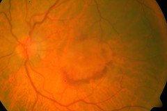
As initially it is a silent disease, mostly it is picked up in a routine retina examination by an ophthalmologist .The retinal examination done by an ophthalmoscope will show the findings of this disease process. To assess the condition in detail, certain other test are done:
Amsler Grid: It is a test paper with graphic picture to be used at reading distance with near glasses on. This is used to check for extent of sight loss-dark spot, distortion or missing of straight lines and also is given to the patient to take home so that he can monitor his symptoms at home and report immediately if there is worsening.
Fluorescin Angiography / ICG: the photographs of macula are taken after injecting a dye in patient’s arm. The dye reaching the eye helps to clarify the type and extent of disease, including detail of abnormal vessels, leaks and membrane formations.
Optical Coherence Tomography : In this test photographs of the retina are taken to show its microscopic detail. So it can help detect any early thickening of the retina in wet ARMD. Also it can delineate the abnormal blood vessels ( choroidal neovascularisation ) from where the blood oozes. Changes of dry ARMD such as drusen can also be demonstrated. Also it is an excellent tool to follow up after treatment to assess the effect of the treatment done and need for re-treatment.
How can it be Prevented ?
There is no prevention of ARMD. Early detection is the key to prevent severe loss of vision. All individuals above 50,especially if there is a family history of ARMD, history of cardiovascular disease, light ocular pigmentation, should get yearly retinal check up for the same. Anyone experiencing following symptoms should consult an ophthalmologist immediately:
Straight lines appearing distorted-specially in the center of vision
Dark blurry or white patch in the center of vision
Color perception changes
Smoking is a risk factor and should be avoided at all costs if any of the risk factor is present.
According to some recent international multicentric trials, multivitamins may slow down progression of dry ARMD. However, excess of fat soluble multivitamins can have their own side-effects and thus consult your doctor before regularly taking multivitamin pills.
How can it be Treated ?
Dry ARMD
There is no permanent cure for dry ARMD. The aim of management is to keep a vigilant check on progression of disease and take measures to improve functional capability of the patient.
· Nutrition: Eat fresh fruits, dark green leafy vegetables. The role of antioxidants/zinc in retarding the progression is not very clear but supplementation with Vitamins A, C and E, zinc and selenium may have a positive effect. A multicentric international trial has demonstrated that Multivitamins slow progression of moderate dry ARMD to severe dry ARMD.
· Sunlight: Blue rays of the spectrum seem to accelerate macular degeneration. Sunglasses with good UV filters for outdoor activities are recommended.
· Smoking: quit smoking as this accelerates the process of ARMD
· Early detection: Monitoring of vision by Amsler grid-report immediately to eye surgeon if any change noticed (the development of wet type may need urgent treatment)
· Low vision aids and lighting –These are devices, which can improve quality of living by improving vision for day-to-day activities, specially reading. Special optical devices like magnifiers (hand held, desktops or in spectacles) can be used in various ways. Adequate lighting will make reading more comfortable with 50-watt indoor bulb in metal shade then fluorescent light.
Wet ARMD
The mainstay of treatment of wet ARMD at present is injection of anti-VEGF drugs into the eye. These are special molecules designed to stop further development of blood vessels. So once the abnormal vessel growth under the retina gets inhibited the leakage of fluid and blood also reduces. However at present these injections need to be repeated at regular intervals as once the effect of the drug wanes off the abnormal vessels star growing again. The two main such drugs being used at present are AVASTIN & LUCENTIS. Presently we donot have a drug which can altogether end the process of this abnormal neovascularisation. A lot of research is ongoing to find a permanent cure for ARMD.
Other treatment modalities available for wet ARMD are -
Photodynamic therapy (PDT) – This involves treating the abnormal vessels with a LASER after injecting a dye which selectively enhances LASER energy absorption by the new vessels only thus preventing damage to the overlying retina. This therapy also may be required to be repeated upto 3 times or more. International studies using this dye have found it to prevent further loss of vision in many cases but it is also not hundred percent effective. Also it does not improve vision but aims to stabilise it, whereas the anti-VEGF injections can improve vision also.
Conventional Laser treatment-This procedure uses a high-energy laser beam to destroy the fragile leaking blood vessels. This will also not improve vision but may reduce further progressive vision loss. However since the high energy laser also destroys retina, it can only be done for lesions away from the central most sensitive part of the retina.
Combinations treatments combining anti-VEGF injections, intraocular steroid injections and PDT are also being tried in some cases.
http://farm4.static.flickr.com/3145/2860280284_6396396a4d_m.jpg
No comments:
Post a Comment